Head Nerve Diagram

Muscular Anatomy Of The Head And Neck With Images Neck Muscle

The Nerves Of The Head And Neck With Images Nerve Anatomy

Scalp Nerve Anatomy Head Find Lots More Of The Best Vintage Book

Cranial Nerves Mnemonic Nerve Anatomy Cranial Nerves Anatomy

Nerves Of Oral Head And Neck Regions Anatomy With Images

Face And Head Nerves With Images Facial Nerve Anatomy Nerve

Nerves of submandibular region.
Head nerve diagram. Epidermis dermis and hypodermis the epidermis is composed of stratified squamous epithelium and is divided into the following five sublayers or strata listed in order from outer. There are 12 of them each named for their function or structure. The sympathetic innervation begins in the spinal cord nerve fibres exit the spinal cord and enter the sympathetic chain which is composed of superior middle and inferior cervical ganglion. The head s blood supply comes mainly from the external and internal carotid arteries.
Ophthalmic maxillary and mandibular. It consists of 15 vector anatomical drawings with 280 anatomical structures labeled. Humeri is a long bone in the arm that runs from the shoulder to the elbow it connects the scapula and the two bones of the lower arm the radius and ulna and consists of three sections the humeral upper extremity consists of a rounded head a narrow neck and two short processes tubercles sometimes called tuberosities. The muscles of the head and neck are also controlled by various cranial nerves including the facial nerve facial expression and accessory nerve head and neck movements.
By passing inferior to the lower border of the superior constrictor of the pharynx and it inclines forwards subsequently at its connection near the posterior end of the mylohyoid line enters the mouth. The humerus ˈ h j uː m ər ə s plural. The vagus nerve is the longest of the 12 cranial nerves. From posterior section of the mandibular nerve the lingual nerve originates and between the ramus of the mandible and the medial pterygoid muscle it descends.
It is intended for the use of medical students working on human anatomy student nurses physiotherapists electro radiological technicians and residents especially those working in neurology neurosurgery otolaryngology and for any physician. Motor cranial nerves help control muscle movements in the head and neck. Instant anatomy is a specialised web site for you to learn all about human anatomy of the body with diagrams podcasts and revision questions. Wandering through the neck and torso the vagus nerve communicates vital information from the brain to the heart and intestines.
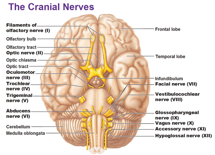
The nerves of the head include the sympathetic and parasympathetic innervation to the head and neck as well as the three branches of the trigeminal nerve. This human anatomy module is about the cranial nerves. Your cranial nerves are pairs of nerves that connect your brain to different parts of your head neck and trunk. The functions of the cranial nerves are sensory motor or both.

Cranial Nerve 7 Facial Nerve Facial Nerve Dental Anatomy

File 1320 The Cranial Nerves Jpg Wikimedia Commons Cranial
The 12 Cranial Nerves With Images Cranial Nerves Vagus Nerve

Brain Stem Anatomy Dengan Gambar

Muscular Anatomy Of Head And Neck Neck Muscle Anatomy Muscle
Cranial Nerve Nuclei In Brainstem Schema 2 With Images

Heart Head And Neck Circulatory System The Cardiovascular System

Brain Anatomy Diagrams With Images Brain Diagram Brain
